baby chest x ray exposure
The chest radiograph is one of the most commonly requested radiographic examinations in the assessment of the pediatric patient. This brochure is to help you understand the issues concerning x-ray exposure during pregnancy.
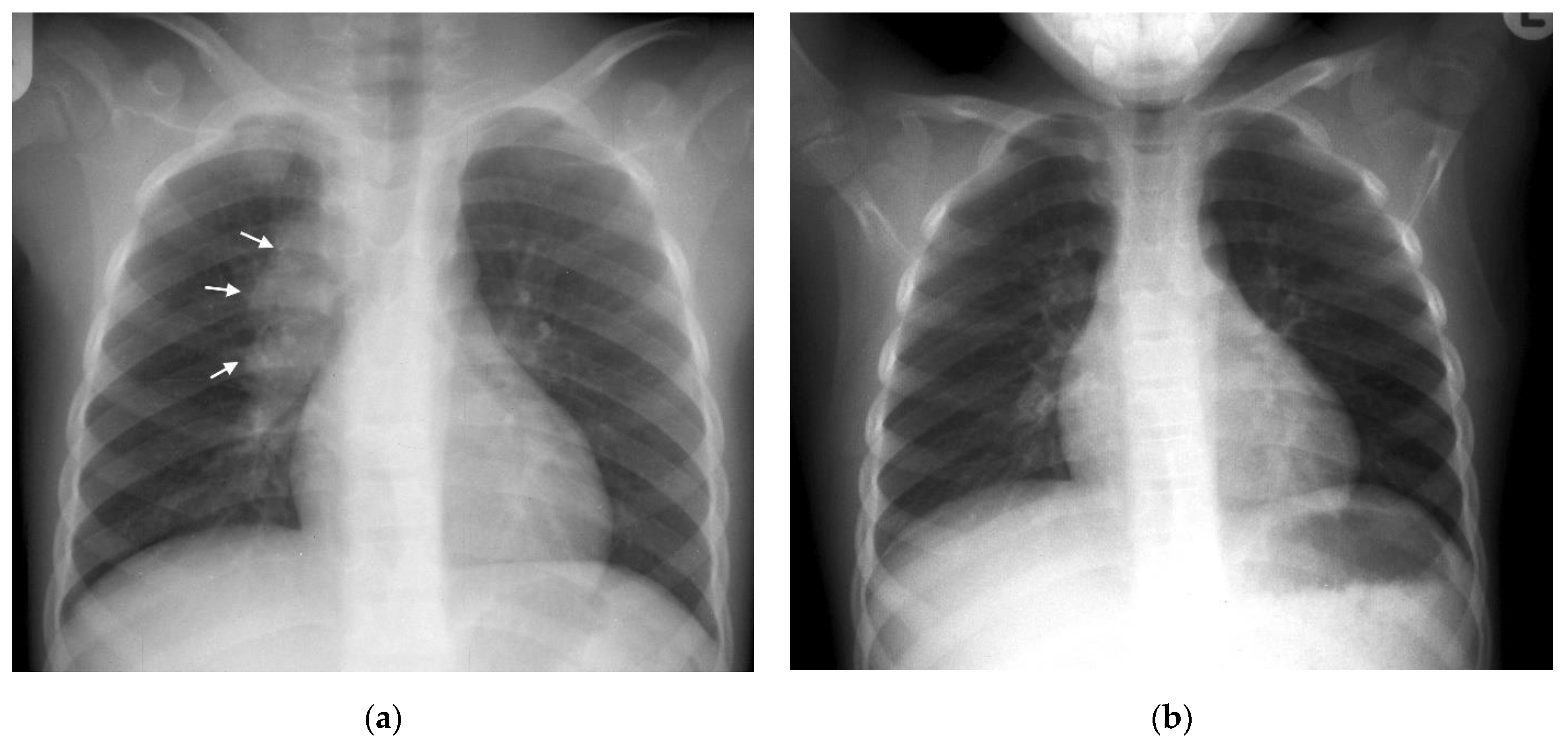
Pathogens Free Full Text Chest Imaging For Pulmonary Tb Mdash An Update Html
Full legfull spine imaging is performed at 180 cm using CR.
. An X-ray beam is absorbed differently by different structures in the body. Risks depend on the amount of radiation to which the baby was exposed and the amount of time that it was exposed. A diagnostic X-ray of your abdomen will expose your baby to slightly more.
Because they spin around the body taking multiple images CT scans can deliver radiation doses that are up to 200 times higher than an. It would take more than 50 chest x-rays to reach 5 rad. A chest X-ray can help doctors.
A child who has had a few X-rays may not have any higher risk. In this study the exposure technique of 65 kVp and 16 mAs was chosen as a reference image due to this technique being near the suitable exposure uses in pediatric chest supine AP in the DR and. The risk of harm to your baby depends on your babys gestational age and the amount of radiation exposure.
It is critical that a pulmonary embolism be ruled out. Erect chest X-rays are taken at 180 cm. The most important characteristic of x rays that concerns us is the.
Exposure to extremely high-dose radiation in the first two weeks after conception might result in a miscarriage. With dental x-rays there is hardly any exposure to any part of the body except the teeth. About one in eight scans ordered for kids is a CT scan.
Imaging should only include areas needing to be evaluated. The scatter that might reach the embryo would be extremely small and would not represent an increased risk for birth defects or miscarriage to your embryo. You explain to your patient that many physicians and patients are unaware of the relatively high baseline risks for adverse outcomes in all pregnancies with or without diagnostic x-ray exposure for major malformations 3 and fetal growth restriction 4.
Since the X-ray beam isnt pointed directly at your abdomen during an X-ray of your teeth chest or arms and legs your baby is exposed to miniscule amounts of radiation youd need to get thousands of X-rays to reach a radiation level that could harm your baby. Exposure to high-dose radiation two to eight weeks after conception might. The lowest amount of radiation possible should be used based on the size of the child.
Imaging should only be used if there is a definite medical benefit. The Alliance for Radiation Safety in Pediatric Imaging reminds parents and pediatricians to follow these guidelines. Diagnostic x-rays can give the doctor important and even life-saving information about a.
When you receive a diagnostic x ray of your chest at a qualified facility the x-ray exposure is not to your embryo. All distal extremity exposures are taken at 110115 cm SID. For example if the radiation dose to the unborn baby was roughly equivalent to 500 chest x-rays at one time the increase in lifetime cancer risk would be less than 2 above the normal lifetime cancer risk of 40 to 50.
Radiation exposure from X-rays may slightly raise the risk of later cancer especially in children who have had many tests with high radiation exposure. It is very rare for a single diagnostic x-ray to exceed even 5 rad. Radiation exposure from X-rays does not pose any short-term problems.
National Center for Biotechnology Information. The scatter radiation that might reach the baby would be extremely small and would not represent an increased risk for birth defects or. When you receive a diagnostic x-ray study of your head teeth chest arms or legs at a qualified facility the x-ray exposure is not to your baby.
X-ray examinations on the arms legs or chest do not expose your reproductive organs to the direct beam. However x-rays of the torso such as the abdomen stomach pelvis lower back and kidneys have a greater chance of exposure to the uterus. However these dose levels arent used in diagnostic imaging.
X-rays are often used to diagnose bone fractures and dislocated joints. A child who has had many CT scans is at greater risk of future cancer. For a simple baby hand xray the child can usually be scanned using a standard device using just a light restraint.
For most children radiation exposure to X-rays probably only raises their risk of cancer a very small amount if at all. Often smaller mini detectors are used for the neonate chest x-ray. A ventilation-perfusion scan would expose the.
51-370 millirad for x-rays of the hip and femur thighbone It is very rare for a single diagnostic x-ray to exceed even 5 rad. Lateral cervical spines are taken at 150 cm. Long-term problems are very small.
The chance of getting cancer increases with the amount of radiation exposure. You would have to x-ray your arm or leg more than 5000 times in order to reach 5 rad of exposure to your unborn baby. Depending on the patients age the difficulty of the examination will vary often requiring a specialist trained radiographer familiar with a variety of distraction and immobilization techniques.

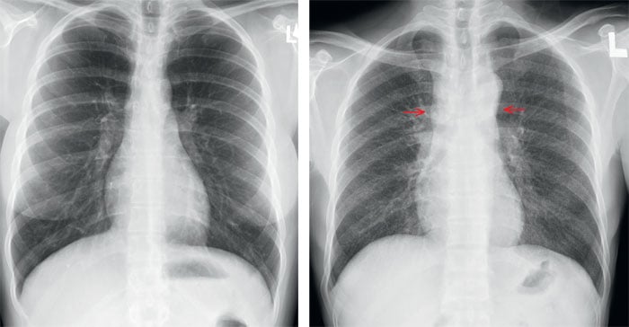
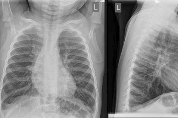
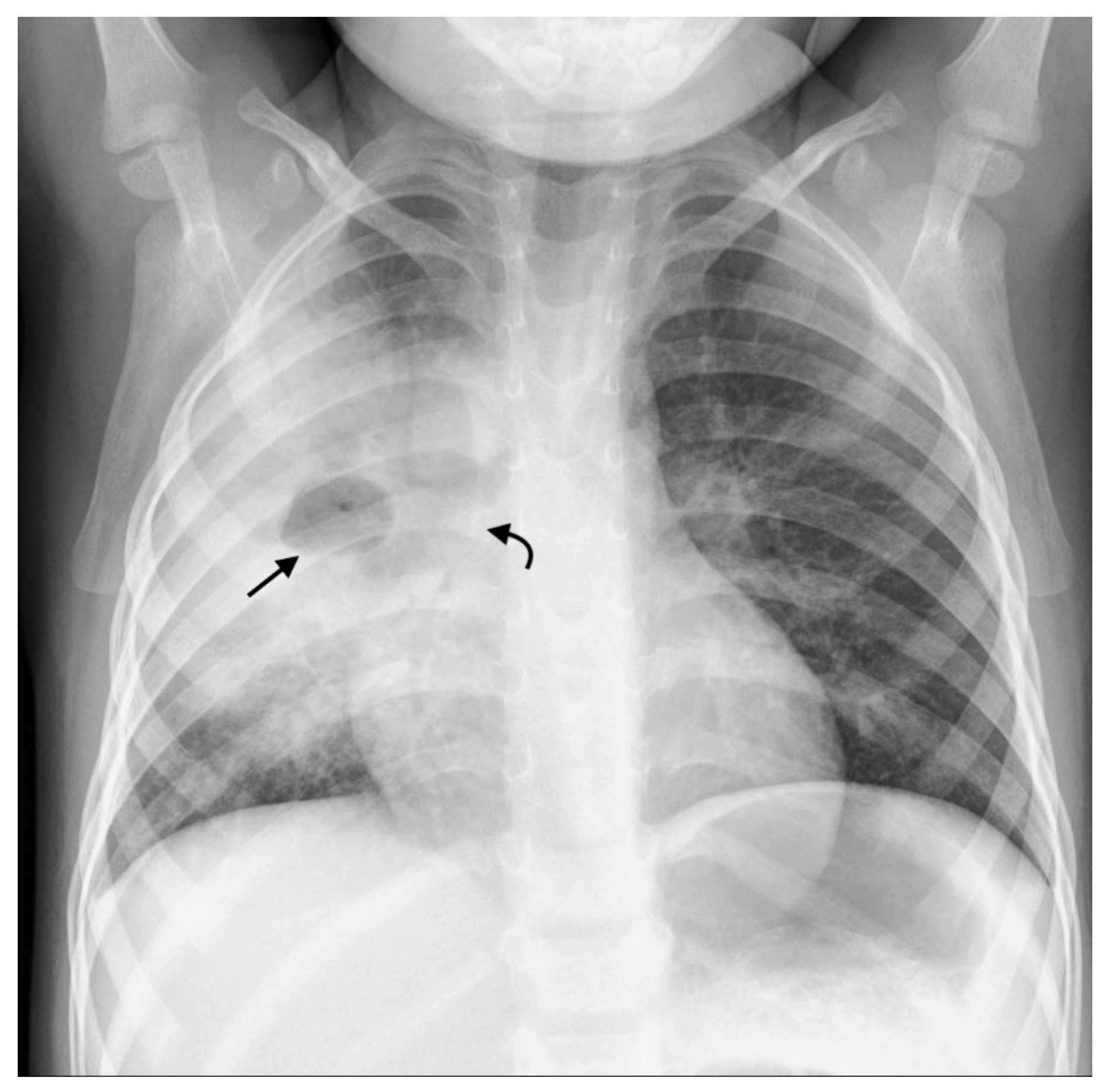
Chest X Ray Lung Imaging Features In Pediatric Covid 19 And Comparison With Viral Lower Respiratory Infections In Young Children Nino 2021 Pediatric Pulmonology Wiley Online Library

Cxr Pediatric I Springerlink

X Ray Imaging For Covid 19 Patients

X Ray Imaging For Covid 19 Patients
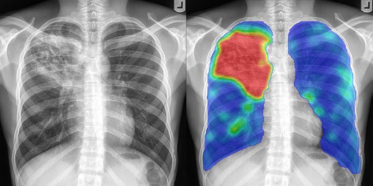
Chest X Ray Analysis Diagnostic Image Analysis Group
Simple Diagnostic Model For Pneumonia In Kids To Reduce Need For X Rays Imaging Technology News
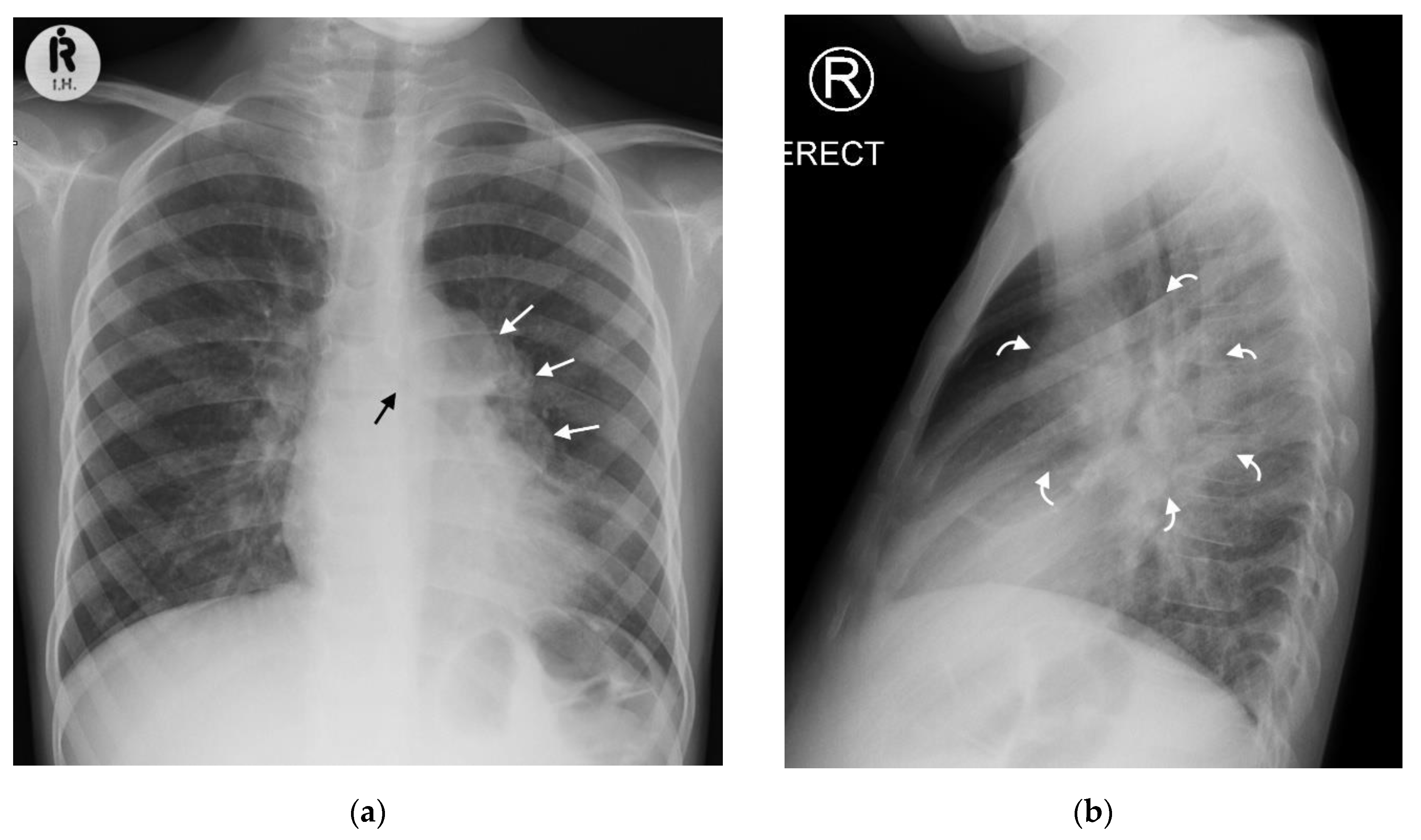
Pathogens Free Full Text Chest Imaging For Pulmonary Tb Mdash An Update Html
Choosing Wisely At Sickkids Means Holding Off On X Raying Infant Bronchiolitis Cases Coming To The Ed

Reliability Of Chest Radiograph Interpretation For Pulmonary Tuberculosis In The Screening Of Childhood Tb Contacts And Migrant Children In The Uk Clinical Radiology

X Ray For Kids Children S Health Orange County

X Ray Imaging For Covid 19 Patients

X Ray Imaging For Covid 19 Patients

Interpretation Of Neonatal Chest Radiography

X Ray Imaging For Covid 19 Patients
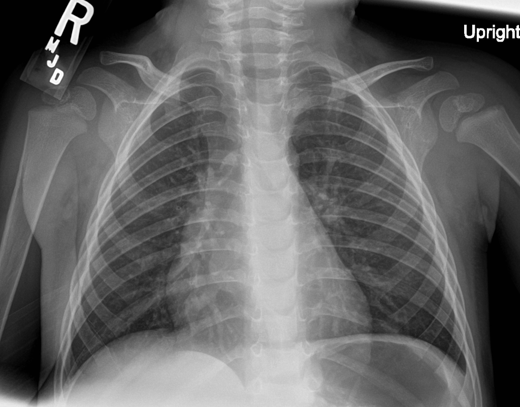
Underinspiration And Poor Positioning Mimicking Lung Pathology In A Pediatric Patient Radiology Case Radiopaedia Org

What Is An X Ray For Kids Radiology And Medical Imaging

Pediatric Chest Supine View Radiology Reference Article Radiopaedia Org
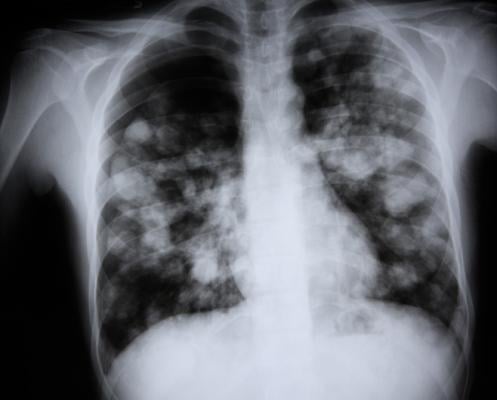
21 Year Old Male Patient With Fever And Hemoptysis Chest X Ray Interpretation
Pathogens Free Full Text Chest Imaging For Pulmonary Tb Mdash An Update Html